animal cell electron microscope
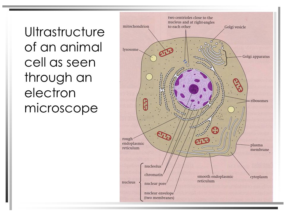
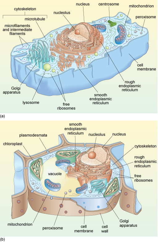
The figure below is a fine structure of a generalized animal cell as seen under an electron microscope. An animal cell also contains a cell membrane to keep all the organelles and cytoplasm contained but it lacks a cell wall.

Cell Lab
What microscope is used to view animal and plant.
. A typical animal cell is 1020 μm in diameter which is about one-fifth the size of the smallest particle visible to the naked eye. Tissue culture cell line infected with human immunodeficiency virus HIV HIV particles are 90-120nm in diameter. This is most likely.
The process of infection. An animal cell also contains a cell membrane to keep all the organelles and cytoplasm contained but it lacks a cell wall. Two types of electron microscopytransmission and scanningare widely used to study cells.
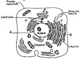
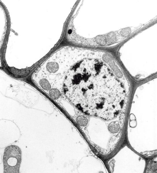
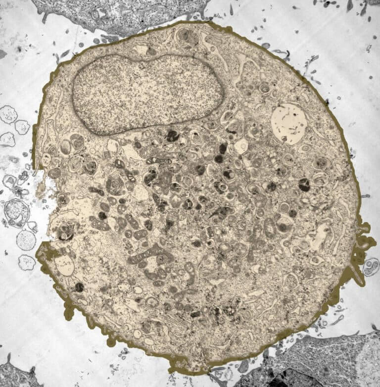
Transmition Electron microscopy of an epithelial cell where mitochondria of several sizes can be seen with the characteristic double membrane and internal cristaes. Animal cells have a basic structure. Up to 20 cash back You can use this royalty-free photo Electron microscope images of animal cells with nucleus and organelles for personal and commercial purposes according to.
We use microscope comprehensively in. How is it different from an animal cell. Beneath a plant cells cell wall is a cell membrane.
Solution Plant cell under an electron microscope. It was not until good light microscopes became. Below the basic structure is shown in the same animal cell on the left viewed with the light microscope and on the right with the transmission.
Observing a wide range of biological processes and animal cell under light microscope is easier due to advances in microscopic techniques. Light and electron microscopes allow us to see. Diagram of animal cell under electron microscope.
What microscope is used to view animal and plant cells. Under the microscope plant cells look like massive rectangular interlocking bricks. A identify cell structures including organelles of typical plant and.
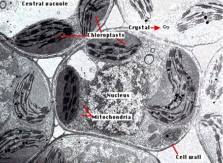
The cell wall of. 1 virus attaches to the cell via. When looking at plant and animal cells with an electron microscope you notice that the plant cells have more Golgi membranes than the animal cells.

Cell Upper Sec Science
Structure Of Plant And Animal Cells Under An Electron Microscope Ppt Video Online Download
The Figure Below Is A Fine Structure Of A Generalized Animal Cell As Seen Under An Electron Microscope

Animal And Plant Cells Gidemy Class Notes

Gce Cie Biology Animal And Plant Cell Structures And

45 Best Cell Diagram Ideas Cell Diagram Cell Plant Cell
Structure Of Animal Cell And Plant Cell Under Microscope Diagrams

Electron Microscopic Structure Of Plant Cell And Animal Cell Brainly In

Cell Micrographs Bioninja

Illustrate Only A Plant Cell As Seen Under Electron Microscope How Is It Different From Animal Cell
Electron Micrographs

Transmission Electron Micrograph Of An Animal Cell Stock Image G450 0052 Science Photo Library
Rem Tem Images Of Plant And Animal Cells Hampden Academy Biology 2012

Biology Notes For A Level 4 Cell Structure And Function
![]()
Electron Micrograph Animal Cell Hi Res Stock Photography And Images Alamy

1 2 Difference Between Plant And Animal Cells Cells As The Basic Units Of Life Siyavula
Amazing 27 Things Under The Microscope With Diagrams
A Tour Of The Cell View As Single Page
The Plant Cell